1. Aduana-Alcantara A.A., Almadrones-Reyes K.J. and Dagamac N.H.A. 2023. Mangrove land suitability assessment using weighted linear combination: A case study of La Union Province Coastline, Philippines. J. Sustainability Resour. Manage. 2 (2): 83-91.
2. Aguilar M. and Lado C. 2012. Ecological niche models reveal the importance of climate variability for the biogeography of protosteloid amoebae. ISME J. 6 (8): 1506-1514. DOI: 10.1038/ismej.2012.12
3. Aguilar M., Spiegel F.W. and Lado C. 2011. Microhabitat and climatic preferences of protosteloid amoebae in a region with a Mediterranean climate. Microb. Ecol. 62: 361-373. DOI: 10.1007/s00248-011-9843-6
4. Balaoro-Banzuela R.C., Ocenar-Bautista C.E., Buebos-Esteve D.E., Claudio-Paragas C.Y. et al. 2023. Rapid diversity assessment of litter myxomycete assemblages in the upland and coastal terrains of San Fernando City, La Union, Philippines. Biodiversitas. 24 (5): 2877-2886. DOI: 10.13057/biodiv/d240542
5. Bernardo J.L.M., Arioder L.J.Q., Almadrones-Reyes K.J. and Dagamac N.H.A. 2018. Myxomycete communities occurring in fragmented forest patches in two municipalities of Laguna, Philippines.Community Ecol. 19: 289-299. DOI: 10.1556/168.2018.19.3.10
6. Buisan P.N.-H.N. and Dagamac N.H.A. 2021. Elucidating hidden slime mold diversity in Southeast Asia: A review of potential methods. Slime Molds. 1: V1A3. DOI: 10.5281/zenodo.5097135
7. Buot I.E., Jr., Origenes M.G. and Obeña R.D.R. 2022. Conservation status of native mangrove species in the Philippines. J. Wetlands Biodivers. 12: 51-65.
8. Cavalcanti L.H., Damasceno G., Costa, A.A.A. and Bezera A.C.C. 2016. Myxomycetes in Brazilian mangroves: species associated with Avicennia nitida, Laguncularia racemosa and Rhizophora mangle. Mar. Biodivers. Rec. 9 (31). DOI: 10.1186/s41200-016-0035-4
9. Dagamac N.H.A. and dela Cruz T.E.E. 2015. Myxomycete research in the Philippines: Updates and opportunities. Mycosphere. 6 (6): 784-795. DOI: 10.5943/mycosphere/6/6/12
10. Dagamac N.H.A., dela Cruz T.E.E., Rea-Maminta M.A.D., Cruz J.A.D. et al. 2017. Rapid assessment of myxomycete diversity in the Bicol Peninsula, Philippines. Nova Hedwigia. 104 (1-3): 31-46. DOI: 10.1127/nova_hedwigia/2015/0252
11. Dagamac N.H.A., Rea-Maminta M.A.D., Batungbacal N.S., Jung S.H. et al. 2015. Diversity of plasmodial slime molds (myxomycetes) in coastal, mountain, and community forests of Puerto Galera, Oriental Mindoro, the Philippines. J. Asia-Pac. Biodivers. 8 (4): 322-329. DOI: 10.1016/j.japb.2015.10.004
12. Dagamac N.H.A., dela Cruz T.E.E., Pangilinan M.V.B. and Stephenson S.L. 2011. List of species collected and interactive database of myxomycetes (plasmodial slime molds) for Mt. Arayat National Park, Pampanga, Philippines. Mycosphere, 2 (4): 449-455. DOI: 10.1080/21501203.2011.637088
13. de Basanta D.W. and Estrada-Torres A. 2017. Techniques for recording and isolating Myxomycetes. In: Myxomycetes biology, systematics, biogeography and ecology (Eds: Rojas C. and Stephenson S.L.). Academic Press, pp. 325-376. DOI: 10.1016/B978-0-12-824281-0.00010-5 EDN: CHQMCU
14. Duke N.C., Ball M.C. and Ellison J.C. 1998. Factors influencing biodiversity and distributional gradients in mangroves. Global Eco. Biogeo. Letters 7: 27-47. DOI: 10.2307/2997695
15. Garcia K., Malabrigo P. and Gevaña D. 2014. Philippines’ mangrove ecosystem: status, threats and conservation. In: Mangrove ecosystems of Asia (Eds: Faridah-Hanum I., Latiff A., Hakeem K., Ozturk M.). Springer. DOI: 10.1007/978-1-4614-8582-7_5
16. Hughes A. 2010. Disturbance and diversity: an ecological chicken and egg problem. Nature Educ. Knowledge. 3 (10): 48.
17. Iwamoto Y. and Nakayama T. 2024. Nannostelium ampullaceum gen. et sp. nov., a tiny new member of the protosteloid amoeba of the Cavosteliida (Variosea, Amoebozoa). Protist. 126046. DOI: 10.1016/j.protis.2024.126046
18. Iwamoto Y., Degawa Y., and Nakayama T. 2023. Reexamination of a rare protosteloid amoeba Schizoplasmodiopsis micropunctata, and the revision of Tychosporium (Cavosteliida, Variosea, Amoebozoa). Mycoscience. 64 (2): 63-68. https://doi.org/10.47371%2Fmycosci.2023.01.002.
19. Jayatissa L.P., Dahdouh-Guebas F. and Koedam N. 2002. A revision of the floral composition and distribution of mangroves in Sri Lanka. Bot. J. Linnaean Soc. 138: 29-43. DOI: 10.1046/j.1095-8339.2002.00002.x
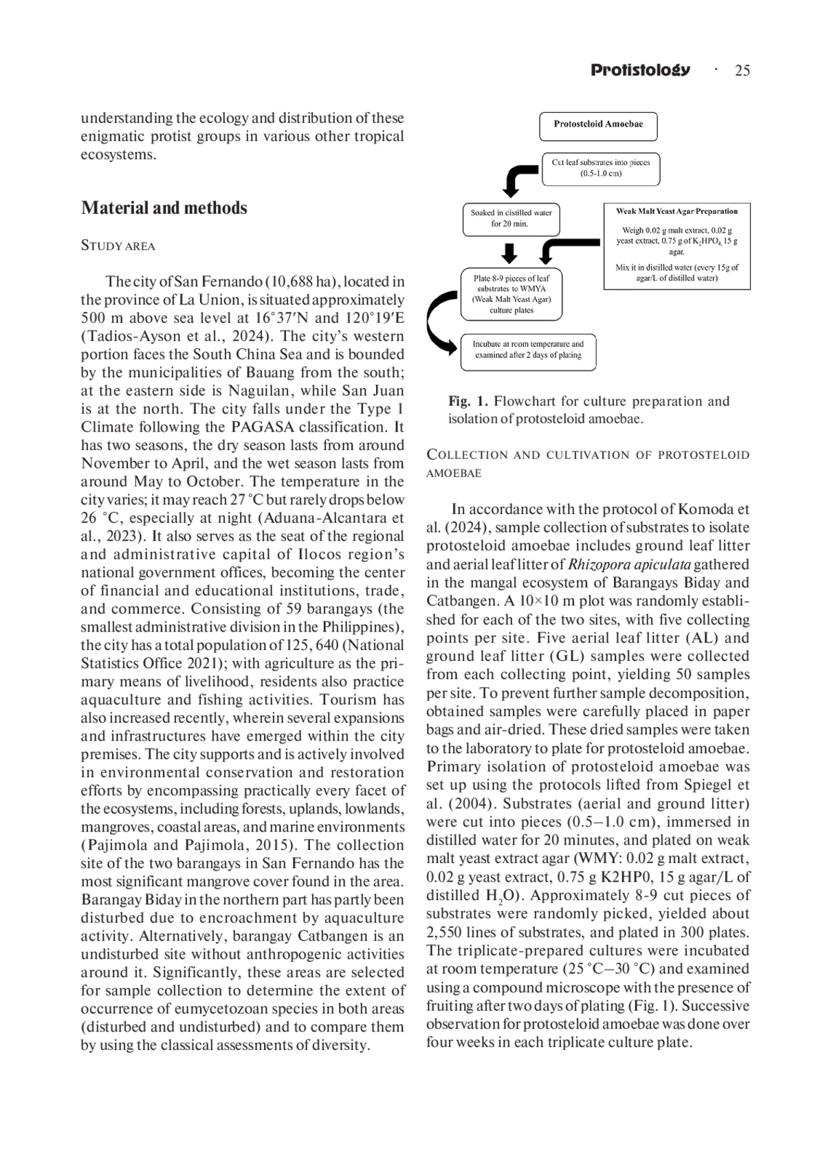
20. Komoda A.T., Tan L.A.D.A, Tomimbang A.M.G., Tan K.A.R. et al. 2024. Protosteloid amoebal assemblages as microbial models for elevational diversity gradient in tropical montane landscape. Sydowia. 77: 129-139. DOI: 10.12905/0380.sydowia77-2025-0129
21. Kudryavtsev A., Brown M.W., Tice A., Spiegel F.W. et al. 2014. A revision of the order Pellitida Smirnov et al., 2011 (Amoebozoa, Discosea) based on ultrastructural and molecular evidence, with description of Endostelium crystalliferum n. sp. Protist. 165 (2): 208-229. DOI: 10.1016/j.protis.2014.02.003
22. Lado C. 2005-2025. An on line nomenclatural information system of Eumycetozoa. http://www.nomen.eumycetozoa.com. Accessed May 14, 2024.
23. Lado C. and Eliasson U. 2022. Taxonomy and systematics: current knowledge and approaches on the taxonomic treatment of Myxomycetes. In: Myxomycetes: biology, systematics, biogeography and ecology (Eds: Rojas C. and Stephenson S.L.). Academic Press, pp. 325-376. DOI: 10.1016/B978-0-12-824281-0.00010-5
24. Lim A., Silva R.M., Lesaca G.R., Mapalo V.J. et al. 2021. First survey of myxomycetes in successional and mangrove forests of Negros Oriental, Philippines. Slime Molds 1: V1A7. DOI: 10.5281/zenodo.5098397
25. Liu Q.S., Yan S.Z. and Chen S.L. 2015. Species diversity of myxomycetes associated with different terrestrial ecosystems, substrata (microhabitats) and environmental factors. Mycol. Progress 14 (27). DOI: 10.1007/s11557-015-1048-9
26. Limbo-Dizon J.E., Almadrones-Reyes K.J., Macabago S.A.B. and Dagamac N.H.A. 2022. Bioclimatic modeling for the prediction of the suitable regional geographical distribution of selected bright-spored myxomycetes in the Philippine archipelago. Biodiversitas. 23 (5): 2285-2294. DOI: 10.13057/biodiv/d230506 EDN: AQTRSB
27. Macabago S.A.B., Dagamac N.H.A., dela Cruz T.E.E. and Stephenson S.L. 2017. Implications of the role of dispersal on the occurrence of litter-inha-biting myxomycetes in different vegetation types after a disturbance: a case study in Bohol Islands, Philippines. Nova Hedwigia. 104 (1-3): 221-236. DOI: 10.1127/nova_hedwigia/2016/0391
28. Macabago S.A.B., Dagamac N.H.A. and dela Cruz T.E.E. 2010. Diversity and distribution of plasmodial myxomycetes (slime molds) from La Mesa Ecopark, Quezon City, Philippines. Biotropia 17: 51-61.
29. Macintosh D.J. and Ashton, EC. 2002. A review of mangrove biodiversity conservation and management. Centre for Tropical Ecosystems Research, University of Aarhus, Denmark.
30. Moore D.L. and Stephenson S.L. 2003. Microhabitat distribution of protostelids in a tropical wet forest in Costa Rica. Mycologia. 95 (1): 11-18. DOI: 10.2307/3761956
31. National Statistics Office. 2021. Census of Population; NSO: Manila, Philippines. https://psa.gov.ph.
32. Ndiritu G.G., Stephenson S.L. and Spiegel FW. 2009. First records and microhabitat assessment of protostelids in the Aberdare Region, Central Kenya. J. Eukaryotic Microbiol. 56: 148-158. DOI: 10.1111/j.1550-7408.2008.00382.x
33. Nguyen L.T.T., Sanchez-Mahecha O., Alma- drones-Reyes K.J., Redeña-Santos J.C. et al. 2019. Occurrence of leaf litter inhabiting myxomycetes from lowland forest patches of Northern and Central Vietnam. Trop. Ecol. 60: 495-506. DOI: 10.1007/s42965-020-00059-9
34. Novozhilov Y.K., Shchepin O.N., Alexandrova A.V., Popov E.S. et al. 2018. Altitudinal patterns of diversity of myxomycetes (Myxogastria) across tropical forests of Southern Vietnam. Protistol. 12 (2): 73-80. https://www.zin.ru/journals/protistology/num12_2/novozhilov_protistology_12-2.pdf.
35. Ocenar-Bautista C.E., Balaoro-Banzuela R.C., Claudio-Paragas C.Y., Buebos-Esteve D.E. et al. 2024. First records of protosteloid amoebae isolated from coastal litter in the Philippines. Check List. 20 (2): 249-257. DOI: 10.15560/20.2.1
36. Ojima M.N. and Jiang L. 2017.Interactive effects of disturbance and dispersal on community assembly. Oikos. 126 (5): 682-691. DOI: 10.1111/oik.03265
37. Pajimola A.H. and Pajimola M. 2015. Environmental sustainable development strategies in the city of San Fernando, La Union. Paper Presented in the 1st Quezon City International Conference, Future Perfect: Cities at the Forefront of Change and Development held at Richmonde Hotel, October 8-9, 2015. www.ssrn.com/abstract=2686049.
38. Polidoro B.A., Carpenter K.E., Collins L., Duke N.C. et al. 2010. The loss of species: mangrove extinction risk and geographic areas of global concern. PLoS ONE 5 (4): e10095. DOI: 10.1371/journal.pone.0010095
39. Powers D.M. and Stephenson S.L. 2006. Protostelids from tropical forests, woodlands and deserts in Australia. Mycologia, 98 (2): 218-222. DOI: 10.3852/mycologia.98.2.218
40. Redeña-Santos J.C., Van Thao D., Schnittler M. and Dagamac N.H.A. 2018. The first report of composition and occurrence of myxomycete assemblages in protected and unprotected plantation forests: a comparative study in Thai Nguyen City, Northern Vietnam. Plant Ecol. Evol. 151 (2): 231-240. DOI: 10.5091/plecevo.2018.1432
41. Shadwick J.D., Silberman J.D. and Spiegel F.W. 2017. Variation in the SSU rDNA of the genus Protostelium leads to a new phylogenetic understanding of the genus and of the species concept for Protostelium mycophaga (Protosteliida, Amoebozoa). J. Eukaryotic Microbiol. 65 (3): 331-344. DOI: 10.1111/jeu.12476
42. Shadwick J.D., Stephenson S.L. and Spiegel F.W. 2009. Distribution and ecology of protostelids in Great Smoky Mountains National Park. Mycologia. 101 (3): 320-328. DOI: 10.3852/08-167
43. Spiegel F.W., Shadwick L.L., Ndiritu G.G., Brown M.W. et al. 2017. Protosteloid amoebae (Protosteliida, Protosporangiida, Cavosteliida, Schizoplasmodiida, Fractoviteliida, and Sporocarpic members of Vannellida, Centramoebida, and Pellitida). In: Handbook of the Protists (Eds: Archibald, J.M., Simpson, A.G.B. and Slamovits, C.H.). Springer, pp. 1311-1348. DOI: 10.1007/978-3-319-32669-6_12-1
44. Spiegel F.W., Shadwick J.D., Lindley L.A., Brown M.W. et al. 2007. A beginner’s guide to identifying the protostelids. University of Arkansas, Fayetteville.
45. Spiegel FW, Stephenson SL, Keller HW, Moore DL et al. 2004. Sampling the biodiversity of mycetozoans. In:. Biodiversity of fungi: inventory and monitoring methods (Eds: Mueller G.M., Bills G. and Foster M.S.). Elsevier Academic Press, pp. 547-576.
46. Tadios-Ayson A.H., Moran C.B. and Dagamac N.H.A. 2024. Modeling invasion patterns of Chromolaena odorata under changing climate and LULC in La Union, Philippines. Vegetos. DOI: 10.1007/s42535-024-01001-3
47. Trierveiler-Pereira L., Baltazar J.M. and Loguercio-Leite C. 2008. Santa Catarina Island mangroves - First report of Myxomycetes on Avicennia schaueriana. Mycotaxon. 103: 145-52.
48. Tomlinson P.B. 1986. The botany of mangroves. Cambridge University Press. DOI: 10.1017/S0266467400002017
49. Walker L.M. and Stephenson S.L. 2016. The species problem in myxomycetes revisited. Protist. 167 (4): 319-338. DOI: 10.1016/j.protis.2016.05.003
50. Zahn G., Stephenson S.L. and Spiegel F.W. 2014. Ecological distribution of protosteloid amoebae in New Zealand. PeerJ 2: e296.